


TBI Research Summary: Cerebellar Network Efficiency Improved by Vielight Intranasal-Transcranial Photobiomodulation (itPBM) (n=30)
This summary details the findings of an n=30 TBI study conducted with the Vielight Neuro RX Gamma, published in Photobiomodulation, Photomedicine, and Laser Surgery (DOI: 10.1177/25785478251376477).
This independent study by the University of Utah (Dr. Wilde et al.) utilized fMRI to detect neurophysiological changes before and after intranasal-transcranial photobiomodulation (itPBM) on ex-football players and athletes suffering from TBI.
Please note: This is research and not medical advice.
Background and Objective
Repetitive head acceleration events (RHAEs) and traumatic brain injuries (TBIs), often sustained in sports or military service, are associated with cumulative neurological compromise. This includes chronic alterations in resting-state functional connectivity (rsFC)—the synchronized activity between brain regions when the brain is not focused on a specific task. While these alterations are documented, few solutions have been investigated to mitigate them.
A previous study by the research team demonstrated that transcranial photobiomodulation (PBM) led to improvements in balance and motor function in adults exposed to RHAEs. The cerebellum is critical for motor function and has extensive connections with cerebral regions targeted by PBM. Therefore, the current study sought to explore the underlying neurological mechanisms by examining changes in the rsFC of the cerebellum following a PBM solution protocol.

Figure 1. Seed connectivity maps. Sample-specific seed-based connectivity maps for each region of interest are overlaid on an array of axial images in MNI template space, lateral–posterior–inferior (LPI) oriented, and represented in neurological view (left = left).
Methodology
- Participants: The study enrolled thirty (30) individuals with a documented history of repetitive head acceleration events.
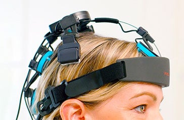
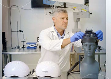
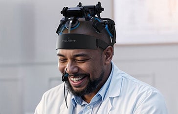
- Intervention: Participants used an at-home transcranial photobiomodulation (PBM) headset, the Vielight Neuro Gamma, which also included an intranasal cannula.
- Protocol: The intervention consisted of 20-minute sessions administered every other day for a period of 8 to 10 weeks.
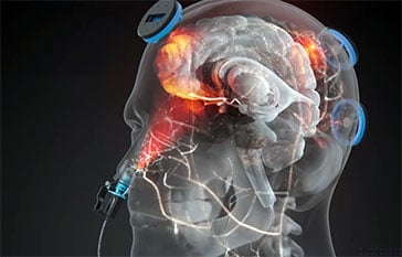
- Dosage: During each session, a total of 240 J/cm² of red and near-infrared light was administered. This light targeted cortical nodes of the Default Mode Network (DMN) as well as subcortical structures.
- Analysis: Resting-state functional magnetic resonance imaging (fMRI) scans were acquired from participants both before and after the 8-10 week stimulation period. The analysis focused on changes in functional connectivity from 11 cerebellar seed regions known to be associated with three major large-scale brain networks:
- The Default Mode Network (DMN)
- The Salience Network (SN)
- The Frontoparietal Network (FPN)

Figure 2. Resting-state functional connectivity (Pre-itPBM) Resting-state functional connectivity (rsFC) of the cerebellum before PBM intervention. Pre > post-treatment rsFC of the default mode, salience, and frontoparietal network seed regions of interest. Peak voxel activity of each associated cluster is overlaid on a template brain standardized in MNI space, lateral–posterior–inferior (LPI) oriented, and represented in neurological view (left = left).
Results
The analysis of the fMRI data revealed significant and specific changes in brain connectivity patterns after Vielight itPBM stimulation:
- Decreased Between-Network Connectivity: Researchers observed an overall decrease in functional connectivity between different brain networks. This suggests a reduction in non-specific or inefficient “crosstalk” between distinct functional systems.
- Increased Within-Network Connectivity: Conversely, the study found an overall increase in functional connectivity within the established networks. This indicates that the connections binding these networks became stronger and more coherent.
- Specific Network Effects: These changes were noted as being particularly prominent within the Salience Network (SN) and the Frontoparietal Network (FPN), two networks crucial for attention, cognitive control, and sensory processin

Figure 3. Resting-state functional connectivity (rsFC) changes (Post-itPBM) Resting-state functional connectivity (rsFC) of the cerebellum after PBM intervention. Post > pre-treatment rsFC of the default mode, salience, and frontoparietal network seed regions of interest. Peak voxel activity of each associated cluster is overlaid on a template brain standardized in MNI space, lateral–posterior–inferior (LPI) oriented, and represented in neurological view (left = left).
Conclusions and Implications
The study’s findings suggest that the Vielight Neuro may improve the “network efficiency” of the cerebellum. The observed pattern of decreased between-network and increased within-network connectivity points toward an increase in “network segregation.”
This study also centers the case around targeted brain photobiomodulation vs pancranial applications, showing that precision and a high-irradiance could lead to potentially superior outcomes by focusing energy where the brain needs to utilize it vs a blanket approach.
In essence, Vielight PBM technology appeared to help the brain regulate overactive, diffuse connections while simultaneously strengthening the integrity of individual, specialized networks. This enhanced organization provides a potential neurological mechanism to explain the improvements in motor function and balance that were reported in the team’s previous work. These results support PBM as a promising noninvasive, at-home therapeutic modality for mitigating the chronic neurological effects of RHAEs.
