
 Shop Vielight tech













Vielight Photobiomodulation Reaches Subcortical Structures: First Human fMRI Evidence
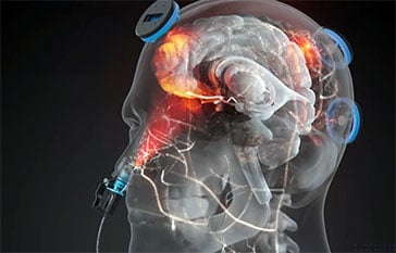
A team at Baycrest put 45 people in an MRI scanner and delivered near-infrared light with Vielight tech through the nose alone. The scanner picked up something forehead-delivered light had not managed in their earlier work: a response in deep brain structures, the thalamus, amygdala and hypothalamus.
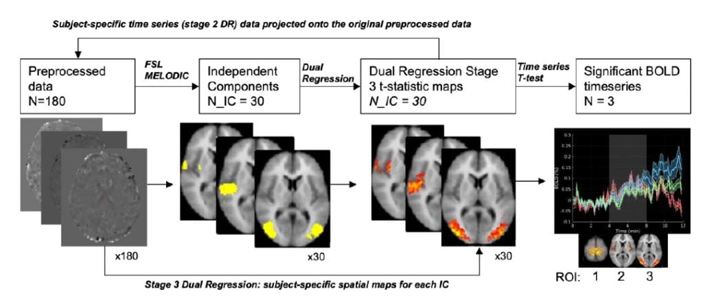
Baycrest Intranasal Study Design (BOLD fMRI Imaging)
Why the nose?
Ask most people where brain light therapy goes and they will point at their forehead. That instinct is reasonable. It is where nearly every device puts its diodes, and it is where nearly every published study has aimed them.
It also carries a hidden assumption: that the shortest path to the brain is a straight line through the front of the head. The skull disagrees. Frontal bone, scalp, and hair scatter and absorb near-infrared light aggressively, and the deeper you want to reach, the less of it survives the trip.
Deep brain structures like the thalamus have long been considered off-limits to non-invasive stimulation for exactly this reason. Transcranial magnetic stimulation cannot reach them directly. Neither can transcranial direct current stimulation.
But there is a second route into the head, and it does not involve going through the skull at all – patented and researched extensively by Vielight.
The cribriform plate
Sitting at the roof of the nasal cavity is a sliver of perforated bone called the cribriform plate. It is the thinnest bone separating the outside world from brain tissue, it is riddled with openings for the olfactory nerve, and it sits directly beneath the orbitofrontal cortex. Light entering through the nostril meets a fraction of the barrier that light entering through the forehead does.
That anatomy has been the theoretical case for intranasal photobiomodulation for years. What has been missing is a study that isolates it. Every previous human trial combined intranasal light with transcranial light, which makes it impossible to say which route did what. A new preprint from the Rotman Research Institute at Baycrest and the University of Toronto is the first to run intranasal delivery on its own inside an fMRI scanner.
Demonstrated energy of Vielight intranasal through a real skull cribriform plate.
Forty-five healthy adults. Four scans each. One hundred and eighty datasets. No forehead light at all.
How the study worked
Participants aged 20 to 32 lay in a 3 Tesla Siemens scanner with an applicator in the right nostril, fed by a 10 metre optical fibre running from a laser console outside the scanner room. They could not see the emitter and were blinded to when it switched on.
Each 12 minute scan followed the same shape: four minutes off, four minutes on, four minutes off. Each participant did four scans with different combinations of wavelength (808 nm or 1064 nm), irradiance (5, 7, or 9 mW/cm²), and pulse frequency (10 Hz or 40 Hz).
The scanner captured two signals at once. BOLD tracks blood oxygenation. Arterial spin labelling tracks cerebral blood flow. Having both allows the researchers to model what the vasculature is actually doing rather than inferring it from oxygenation alone.
One methodological detail matters more than it sounds. The team did not decide in advance which brain regions to examine. They used independent component analysis to let the data surface whichever regions tracked the light’s on/off timing, precisely because nobody yet knows what an intranasal light response is supposed to look like. The regions below were found, not chosen.
What the scanner saw
Five regions emerged with responses locked to the stimulation timing, plus the illumination site itself.
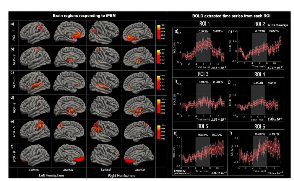
Brain regions responding to Vielight intranasal, tracked through BOLD-fMRI neuroimaging.
Subcortical structures responded
The thalamus and caudate showed a measurable response, as did the amygdala and hypothalamus. This is the finding the authors lead with, because their own earlier transcranial work using the same scanner, the same team, and the same analysis pipeline found no subcortical response at all. The comparison is unusually clean.
Far less light, comparable response
The intranasal protocols ran at 5 to 9 mW/cm². That is a small fraction of typical transcranial irradiance. Normalising response magnitude by power density, the authors calculate that intranasal delivery produced roughly 28 times the response per unit of irradiance. Their phrasing is careful and so is ours: this is an efficiency ratio, not a claim that the absolute effect was larger.
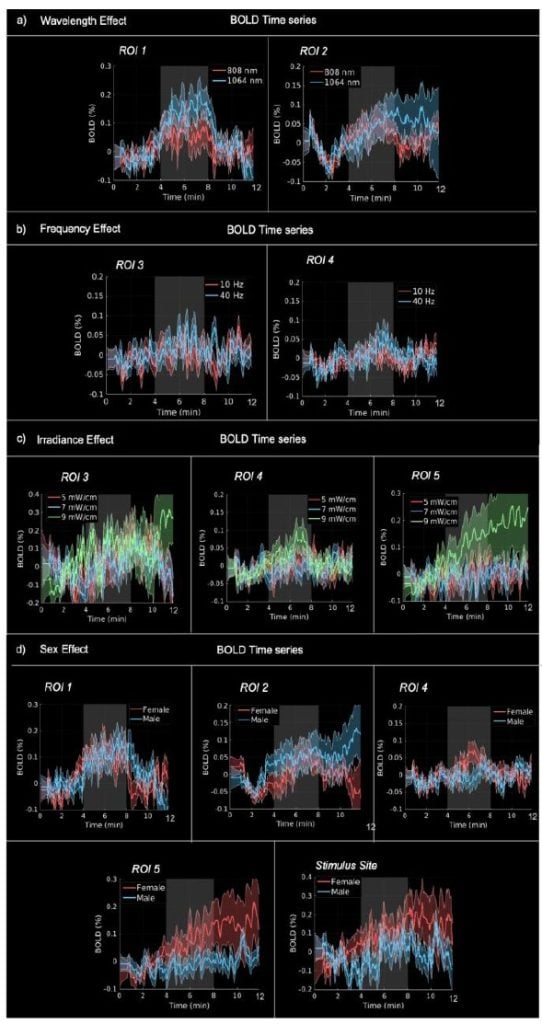
Analysis of BOLD-fMRI signatures.
The response unfolds over minutes, not seconds
Three temporal shapes appeared. Some regions switched on and off with the light. Others climbed steadily and kept climbing after the light stopped. The thalamus and the illumination site did something stranger: they rose during stimulation, dipped when the light went off, then rose again. Across regions, blood flow increased more than metabolic demand required. Whatever is happening, it is not an instantaneous switch.
Sex shaped the response more than expected
Females showed higher responses in the subgenual region, amygdala, parietal cortex, and at the illumination site. Males showed a higher response in the thalamus. These differences were considerably more pronounced than in the same team’s transcranial data, which suggests dose personalisation may matter more for this delivery route rather than less.
Blocked, or bypassed
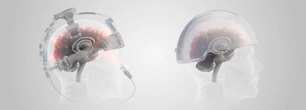
Through the skull
Light meets scalp, hair, and frontal bone. Scattering and absorption take most of it. Cortical regions near the emitter respond. In this team’s data, deeper structures did not. Higher irradiance is needed to compensate, which raises thermal considerations at the skin.
Through the nose
Light enters through the nasal cavity and meets the cribriform plate, the thinnest bone in the path. Cortical regions responded. So did the thalamus and amygdala. It took a fraction of the power density, which the authors note removes thermal effects from the equation entirely.
What this does not show
This is a preprint. It has been posted publicly but has not yet cleared peer review, and its findings should be read as provisional until it does.
The authors are direct about the rest. They cannot say how the light reached those deep structures. Optical propagation through tissue, transmission along cerebrospinal fluid, movement via the olfactory tract, or a vascular route are all candidates, and this study cannot distinguish between them. One of the responding regions, the superior temporal cortex, is the furthest region from the nostril, which is difficult to explain by direct penetration alone.
The exposure was a single four minute session in healthy adults aged 20 to 32. It captures an acute physiological response. It says nothing about what daily use over weeks would do, and nothing about older brains or clinical populations. The absolute signal changes were small, in the range of hundredths of a percent, which is normal for this measurement but worth stating plainly.
And a physiological response is not a clinical outcome. Showing that light changes blood flow in the thalamus is a long way from showing that it changes how someone thinks, feels, or functions. That work has not been done here.
Why it is worth watching
The thalamus routes almost everything. Sensory traffic, motor signals, attention, sleep architecture. It is disrupted across a long list of neurological conditions, and it has been structurally out of reach for non-invasive stimulation. The amygdala sits at the centre of emotional processing and is similarly difficult to target.
Emerging techniques like focused ultrasound are beginning to reach these depths. This preprint suggests near-infrared light delivered through the nose may be another way in, at power densities low enough that heat is not a design constraint.
The question this study opens is not whether the nasal route works. It is why it works better than the physics predicted. Simulations expected around one percent of incident energy to make it through. The measured response outran that prediction, which means something in living tissue, most likely the vasculature, is doing part of the work. That is the next study, not this one.
Study: Van Lankveld H, Chen JX, Zhong XZ, Chen JJ. Intranasal photobiomodulation: an energy efficient paradigm for cortical and subcortical stimulation. bioRxiv preprint, posted March 5, 2026. DOI: 10.64898/2026.03.03.709361. This preprint has not been certified by peer review.
Disclosure: Vielight Inc. supplied the MRI-compatible laser systems used in this study and is acknowledged by the authors as a research partner. The study was funded by the Ontario Centre for Innovation, the Natural Sciences and Engineering Research Council of Canada, and private donation.
Vielight devices are general wellness products. They are not intended to diagnose, treat, cure, or prevent any disease or medical condition, including traumatic brain injury, concussion, chronic traumatic encephalopathy, or any neurological or psychiatric disorder. Research described here is exploratory and does not establish clinical benefit. Consult a qualified healthcare professional regarding any health concern.


