
 Shop Vielight tech











The Brain Network That Breaks First in Alzheimer's
Most people think Alzheimer’s begins with forgetting.
A misplaced name, a forgotten appointment, the word on the tip of the tongue. It is a reasonable assumption. It is also, according to the brain imaging, wrong. By the time memory visibly slips, something else has been coming apart quietly for years.
What Actually Changes First
Research indicates the earliest changes in Alzheimer’s are not about storage. They are about connection. A network of brain regions called the Default Mode Network (DMN), the system that stays busy when your mind is at rest, remembering, imagining, reflecting, begins to lose its coordination well before symptoms surface.
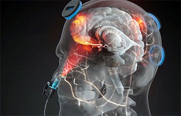
The DMN links the precuneus, posterior cingulate cortex, medial prefrontal cortex, and lateral temporal regions into one coordinated system. In early Alzheimer’s, those links thin out. The network, in effect, starts to fracture. Memory loss is not the beginning of the story. It is what becomes visible once the fracturing is already well underway.
That reframing changes the question worth asking. Not just “can we slow memory loss,” but “can we do anything about the connectivity that fails first?” A new University of Toronto pilot study set out to test exactly that.
A Trial Built to Watch the Network
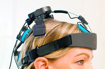
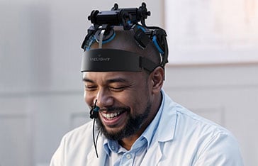
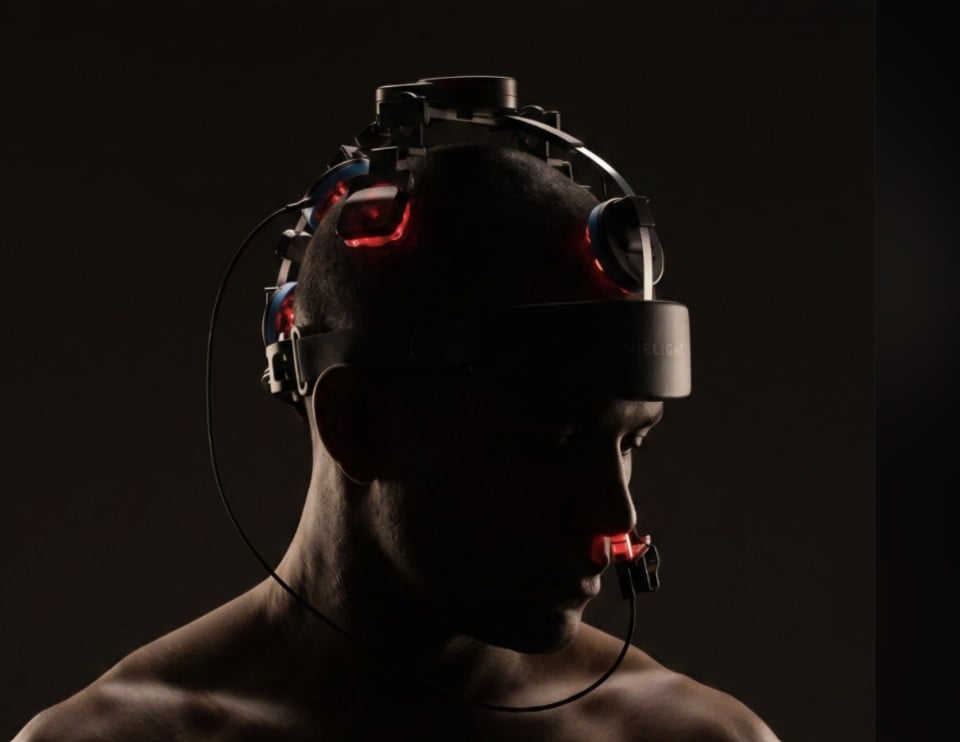
Researchers at the University of Toronto and St. Michael’s Hospital, publishing in the Journal of Alzheimer’s Disease, ran a randomized, sham-controlled multimodal clinical trial in 20 adults with mild cognitive impairment (MCI), the stage where thinking has measurably slipped but independence remains intact. Half used an active Vielight itPBM device six days a week for six weeks. Half used an identical inactive one.
What set the design apart was that it did not measure one thing. It tracked three at once in the same people: brain connectivity on MRI, blood-based metabolism, and cognition. When independent lines of evidence move together, they start to corroborate one another.

Fractured, or Reconnected?
That was the difference the imaging captured between the two groups. On resting-state MRI, the two arms did not look the same at the end of six weeks.
Sham group
Fractured
Memory scores drifted downward over the six weeks, the direction expected as MCI progresses without intervention.
Active itPBM group
Reconnected
DMN connectivity increased versus sham, with a large effect size (d = 1.25, p = 0.014). Memory improved rather than declined.
The network most associated with memory, and one of the first to break down in Alzheimer’s, showed measurable reconnection in the people who received active light. Connectivity between the caudate and the DMN rose as well, consistent with stronger integration of memory and cognitive-control circuits.
And the rest moved with it
Episodic memory (CVLT-II): improved while sham declined (d = 1.09)
Global cognition (MMSE): improved within the clinically meaningful range (d = 1.05)
Brain energy and inflammation: more efficient mitochondrial metabolism, lower IL-6
The connectivity finding did not stand alone. Memory improved. Global cognition improved. Markers of brain energy shifted toward the efficient, mitochondria-driven pathway, and an inflammatory marker tied to cognitive decline fell. The network reconnected, and the wider picture moved with it.
Not the First Time the DMN Network Responded
What makes this harder to dismiss is that the same network keeps showing up across independent imaging studies of Vielight technology, in different populations and at different universities.
At the University of California San Francisco and the San Francisco VA, a pilot in older adults with dementia found increased connectivity between the posterior cingulate cortex and lateral parietal nodes of the DMN, alongside improved cerebral perfusion and better cognitive scores. At the University of Utah, adults with a history of repetitive head impacts showed tighter within-network coherence and less noisy cross-talk after using the device. And at the University of Sydney, healthy young adults showed the flip side of the same regulation: the DMN quieting down appropriately when it was time to focus.
The throughline is not simply more activity. It is normalization, the network behaving as it should for the moment. The MCI trial adds resting-state reconnection in early cognitive decline to that body of evidence, and pairs it, for the first time, with matching changes in memory, brain energy, and inflammation in the same people.
Link: Read the full imaging story across The Universities of Sydney, UCSF, and Utah

Why This Approach Reaches the Network
The device used 810 nm near-infrared light, the wavelength most efficiently absorbed by cytochrome c oxidase, the mitochondrial enzyme central to cellular energy production. Four emitters targeted cortical regions of the DMN, and a fifth intranasal emitter delivered light toward the olfactory bulb, one of the earliest sites of Alzheimer’s-related change and directly connected to the limbic and prefrontal hubs of the network.
This combination of transcranial and intranasal delivery, the intranasal-transcranial (itPBM) configuration at the core of Vielight’s technology, may reach circuits that scalp-based light alone cannot. The network-level effects seen in the trial may partly reflect that route.
Free Clinical Trial Results Webinar
Hear the results directly from the scientist behind the study
In just over a week, Dr. Neda Rashidi-Ranjbar, MD, PhD (University of Toronto), one of the physician-scientists who led this trial, will walk through the findings live and take questions on what the data does and does not show.
July 31, 2026 | 2:00 PM EST
What It Means, and What Comes Next
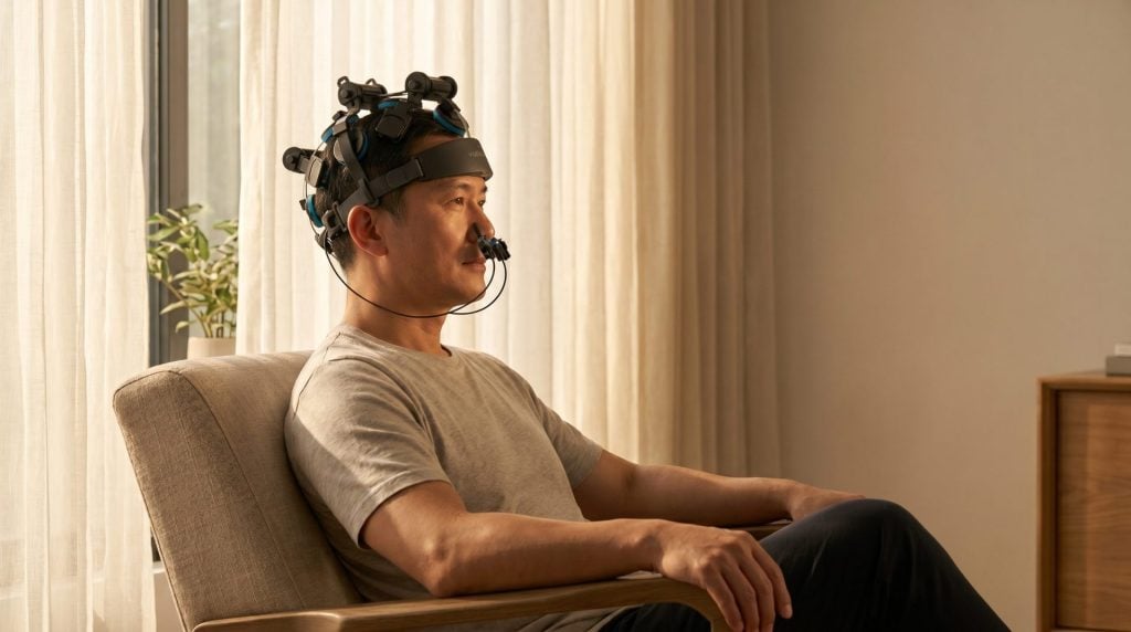
This was a pilot study of 20 people. Large effect sizes in a sample this size signal a strong preliminary result worth confirming, not a settled conclusion. The authors describe home-based itPBM as safe, well tolerated, and feasible, with adherence near 97% and no serious adverse events, and they call for larger, double-blind, multicenter trials.
Still, the direction is notable. If the network fractures first, then a therapy that shows measurable reconnection, alongside gains in memory, brain energy, and reduced inflammation, all from a non-invasive device used at home, is worth understanding in full. And worth hearing about from the researchers themselves.
Read our full breakdown of the MCI trial results
Reference: Rashidi-Ranjbar N, et al. A multimodal evaluation of intranasal-transcranial photobiomodulation in mild cognitive impairment. Journal of Alzheimer’s Disease. 2026.


