
 Shop Vielight tech







Illuminating the Brain: The Vielight Neuro’s Energy Footprint Through a Real Human Skull
Five LEDs don’t make five dots of light. We filmed it through a real human skull.
Point a near-infrared LED at a skull and you might expect a small bright spot on the other side. That is not what happens. Here is what the light actually does on its way to the brain, and why the route in matters as much as the power.
The assumption: one LED, one spot
It is natural to picture light like a laser pointer: a narrow beam that lands on one small patch of brain. If that were true, a headset with five LEDs would reach five small patches and miss everything in between.
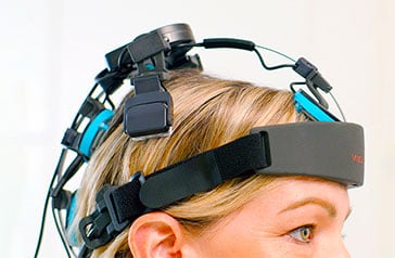
To see what really happens, we placed the Vielight Neuro 4 on a human calvaria (the dome of the skull) and filmed the other side with a camera that can detect 810 nm near-infrared light, which is invisible to the human eye.
Vielight Neuro 4 through a human calvaria. Each LED produces a broad halo, not a point, and the halos overlap.
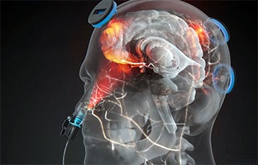
Each LED emerges as a wide halo of light, brightest at the contact point and fading outward. The halos from neighbouring modules overlap, so five modules cover far more of the cortex than five dots ever could.
Why a point becomes a halo
Light reaching the brain passes through four layers, and each one changes its shape.
1. Scalp. Collagen, fat and small blood vessels absorb part of the light and scatter the rest. This is where the biggest losses happen, which is why output power at the surface matters.
2. Skull. Bone is uneven at a microscopic level and scatters light strongly. A narrow beam that enters the bone leaves it as a wide, diffuse halo.
3. Cerebrospinal fluid. The fluid around the brain is clear, so it scatters very little. Light that reaches this layer tends to travel sideways along it before entering the cortex, spreading the halo further over the surface of the brain’s folds.
4. Cortex. Grey matter scatters light mostly forward and absorbs it as it goes. The footprint keeps widening, while intensity falls with depth.
In short: the head is a diffuser. Scattering turns a few powerful points of light into large, overlapping fields, and LED placement decides where those fields concentrate.
Where the halos concentrate: the Default Mode Network
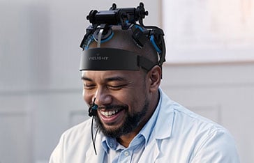
The Neuro 4’s modules are positioned over the main hubs of the Default Mode Network (DMN), the brain network most active during rest, memory recall and self-reflection. It is one of the most studied networks in brain imaging research.

The Default Mode Network in brain scans across different mental states.
Because the halos spread, light still reaches frontal, temporal and lateral parietal areas. The result is broad cortical coverage, weighted toward the DMN.
What light from above can’t easily reach
Scattering explains how light covers the top and sides of the brain. It does not solve the underside. The base of the frontal lobe sits behind thick frontal bone and the sinuses, a long path for light that enters from the scalp.
The nasal cavity offers a shorter one. At its roof is the cribriform plate, a thin, porous bone that sits directly beneath the underside of the frontal lobe. Vielight’s intranasal module delivers light from there.
Light from the Vielight intranasal module, filmed through the skull.
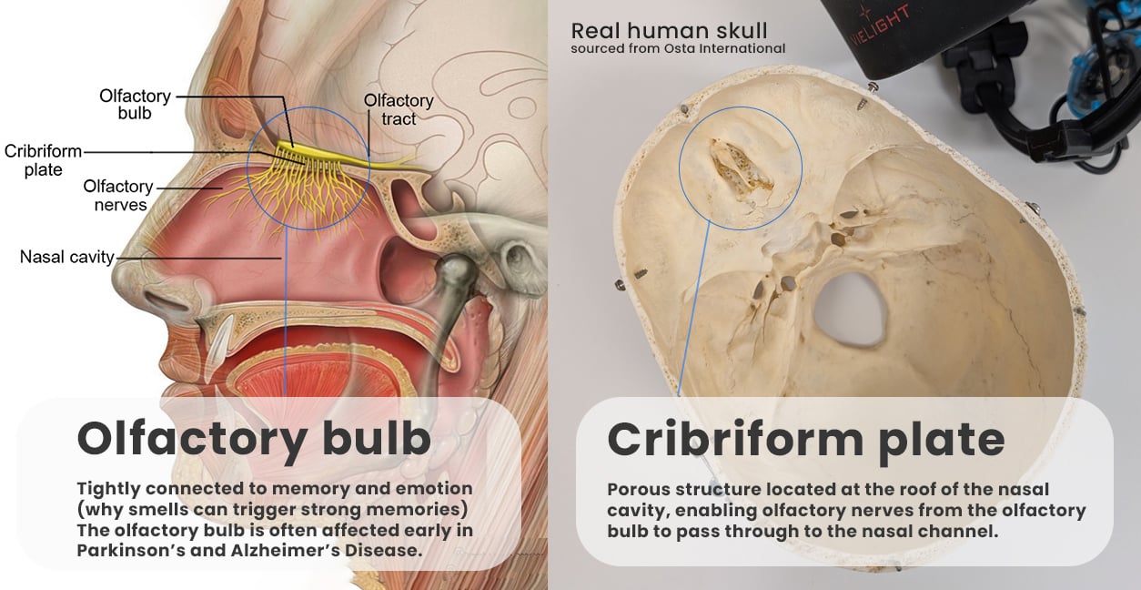
The cribriform plate, the thin bone between the nasal cavity and the brain.
This route puts light close to ventral regions including the orbitofrontal cortex and ventromedial prefrontal cortex, which are involved in emotion regulation and decision-making, and the olfactory bulbs, which handle smell.
The route is efficient. In a 45-person MRI study at Baycrest’s Rotman Research Institute, published in Brain Stimulation in 2025, light delivered at the nostril produced cerebrospinal fluid responses similar to light on the forehead while using about one-twentieth of the power density. The study used research lasers rather than consumer devices, tested each route separately in healthy young adults, and measured single short sessions. Read our summary of the study.
Light from above vs. light from above and below
| Transcranial only (helmet-style devices) |
Intranasal-transcranial (itPBM) (Vielight Neuro) |
|
|---|---|---|
| Light enters from | The scalp | The scalp and the nasal cavity |
| Path to the brain’s underside | Through thick frontal bone and sinuses | Through the thin cribriform plate |
| Regions in the direct light path | Top and sides of the cortex | Top and sides of the cortex, plus ventral frontal regions |
Vielight calls this combination intranasal-transcranial photobiomodulation (itPBM).
More modules, more control: the Neuro Pro 2
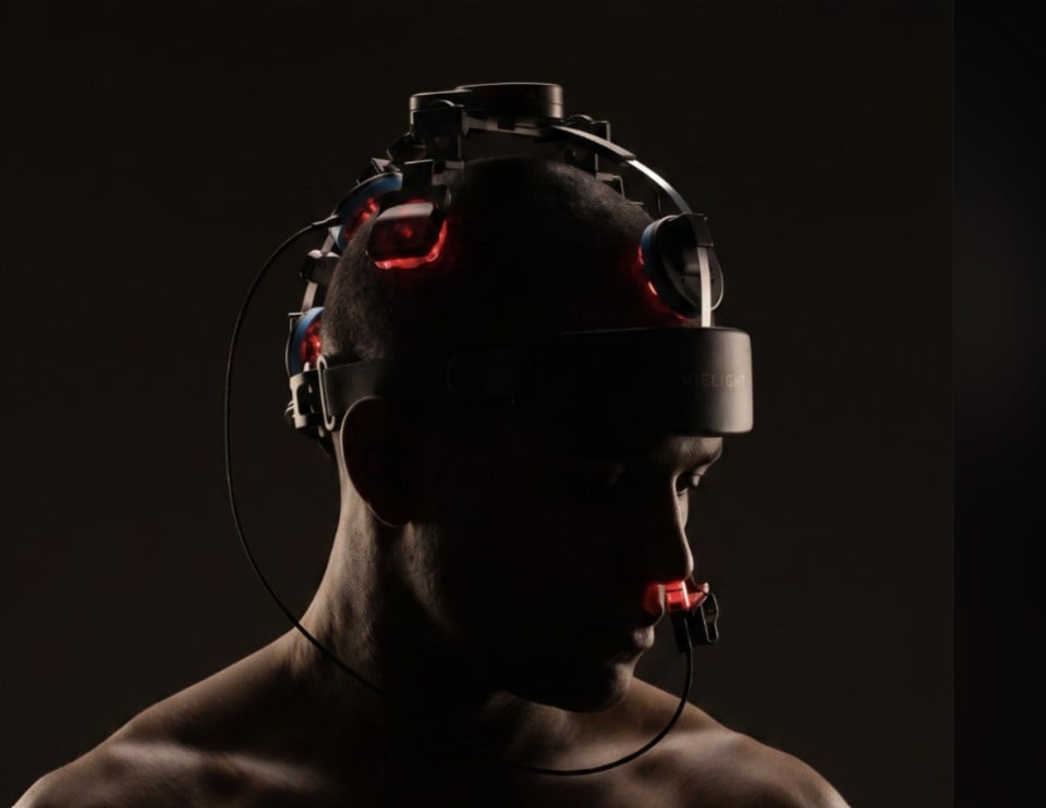
The Neuro Pro 2 applies the same physics with twelve higher-output Vie-LED modules, each individually programmable.
Vielight Neuro Pro 2 at 810 nm, filmed through a human skull.
More light at the surface. Twelve high-output modules put more light into the scalp, where hair and thicker bone cause the largest losses.
Network targeting. Modules can be weighted toward specific networks, such as the DMN or the frontoparietal network.
App control. Nine preset protocols, plus module-level control of frequency, pulsing, irradiance, sweeps and phase.
| Vielight Neuro 4 | Vielight Neuro Pro 2 | |
|---|---|---|
| Vie-LED modules | 5 high-power | 12 higher-power |
| Network emphasis | Default Mode Network | Programmable (DMN, salience, executive and more) |
| Intranasal | Single pathway | Dual pathways |
| Control | Plug-and-play, fixed protocols | Neuro Pro app, fully programmable |
| Best for | Simple daily brain wellness sessions | Performance users, practitioners and researchers |
In the lab: measuring light through skull and skin
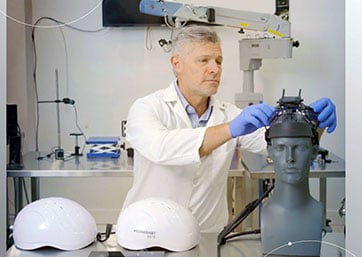
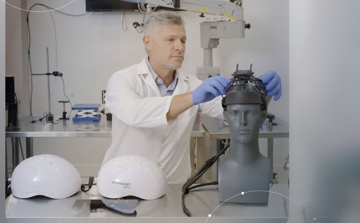
Surface irradiance is only half the story. What matters is how much light is left after the skin and skull. In our photonics lab, we follow the measurement method from the PBM Foundation’s 2024 independent irradiance reports, conducted with Optronic Lab and Megalab, then add the barriers light meets in real life: a human calvaria and a synthetic skin phantom.
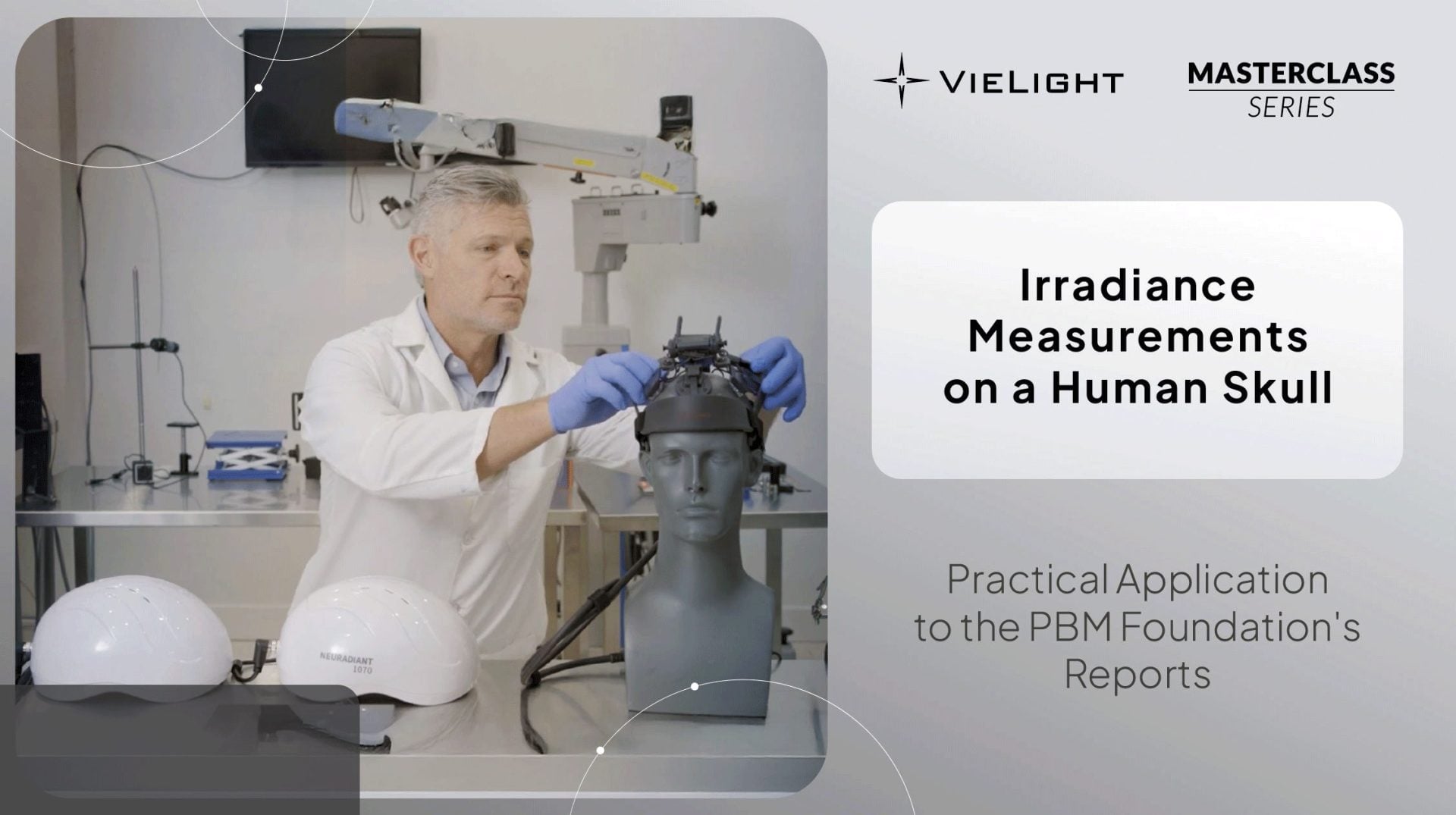
Vie-Lab demo: light penetration through a human skull and skin phantom.
We used the same setup to compare the Neuro Pro 2 with a Neuronic 1070 nm LED helmet. Watch the side-by-side comparison.
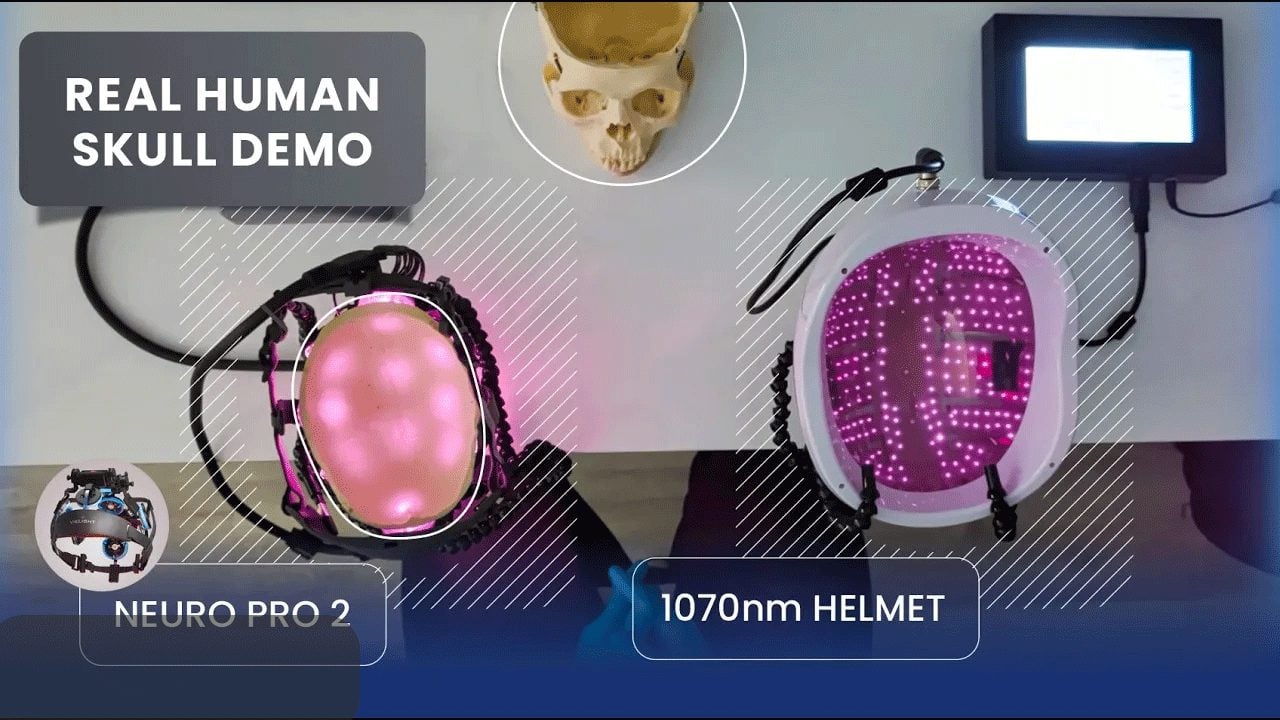
What this footage shows, and what it doesn’t
The videos show the shape and relative intensity of light passing through bone. They are a visualization, not a dose measurement: camera sensitivity and automatic exposure affect how bright the image looks, and the footage was captured on a single skull.
Skull thickness, sinus size and fluid layers vary from person to person, so footprints vary too. Calibrated in-tissue measurements and optical modelling are the tools for quantifying dose, and linking delivered light to biological response remains an active area of research.
Light reaching tissue is a precondition for photobiomodulation. It is not, on its own, evidence of a benefit. For that, see the published research on Vielight technology.
Five points of light, one footprint
Scattering turns a handful of powerful LEDs into broad, overlapping fields across the top of the brain, with placement deciding where they concentrate. The intranasal route adds what light from above struggles to reach: the underside of the frontal lobe, through the thinnest bone between the outside world and the brain.
Explore Vielight Neuro devices
Vielight devices are general wellness products. They are not intended to diagnose, treat, cure, mitigate or prevent any disease or medical condition, including traumatic brain injury, concussion or CTE. Laboratory footage shows light transmission through skull specimens and does not demonstrate health outcomes.


