
 Shop Vielight tech










Brain Imaging Studies with Vielight Neuro Reveal the Network Effects of Brain Photobiomodulation
The misconception: a louder brain is a better brain
It’s tempting to assume that anything good for the brain should make it more active. But a focused brain isn’t the loudest brain. It’s the one that knows which regions to turn up and which to turn down.
Meet the Default Mode Network
The Default Mode Network (DMN) is a set of regions that includes the posterior cingulate cortex, the medial prefrontal cortex, and the lateral parietal areas. It’s most active when your mind is resting, daydreaming, or turned inward. When you switch into a demanding task, it should quiet down so task-focused regions can take the lead.
That switch, from mind-wandering to focus, is a core part of healthy attention.

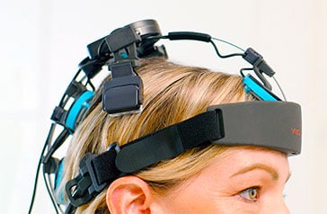
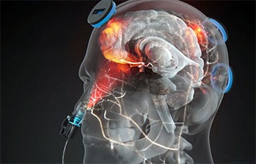
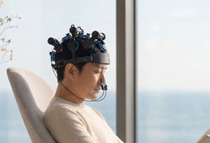
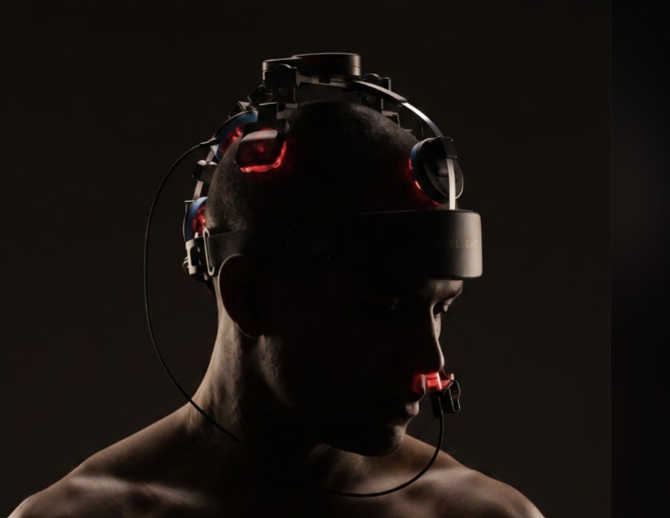
The Vielight Neuro’s geometry is designed so its light fields overlap over key DMN nodes while still covering the wider cortex. That’s how five VieLED modules deliver what is effectively full-transcranial coverage, with an emphasis on the DMN.
Why emphasis on hubs matters
Near-infrared light loses intensity quickly as it passes through the scalp and skull, so how much reaches a region depends on where the light is aimed and how concentrated it is. Spreading light evenly across the whole head sounds thorough, but it means no region gets priority, including the ones that coordinate how the rest of the network behaves.
The DMN is organized around a few highly connected hubs, such as the posterior cingulate cortex, precuneus, and medial prefrontal cortex. Because hubs sit at the crossroads of network communication, they’re a logical place to concentrate light. The Vielight Neuro is built around that idea: overlapping fields put more light on key hubs, broader coverage still reaches the surrounding cortex, and the intranasal module adds a second pathway from beneath. Think of it as lighting a city’s main intersections brightly while keeping every street lit, rather than spreading the same dim glow everywhere.
This describes the design rationale. Research into how light targeting shapes network activity is ongoing.
The study: University of Sydney
El Khoury, H., Mitrofanis, J., & Henderson, L. A. (2019). “Exploring the Effects of Near Infrared Light on Resting and Evoked Brain Activity in Humans Using Magnetic Resonance Imaging.” Neuroscience. Read the study
Who: 24 healthy young adults
What: A 20-minute active session and a 20-minute sham session with a Vielight Neuro Alpha (810 nm, pulsed at 10 Hz), 4 to 12 weeks apart, in random order, without participants knowing which was which. Each session was followed by fMRI at rest and during a simple finger-tapping task.
Independence: No author was affiliated with Vielight, and the researchers bought the device from Vielight’s Australian outlet.
What they found
- At rest: global brain activity didn’t change. The light didn’t simply “rev up” a resting brain.
- During the task: task-evoked activity was lower in the putamen, primary somatosensory cortex, and parietal association cortex.
- Connectivity: one specific shift appeared, in the functional connectivity of the parietal association cortex.
The researchers linked these changes to attention and novelty. They noted that the pattern “served to deactivate the so-called default mode network.”
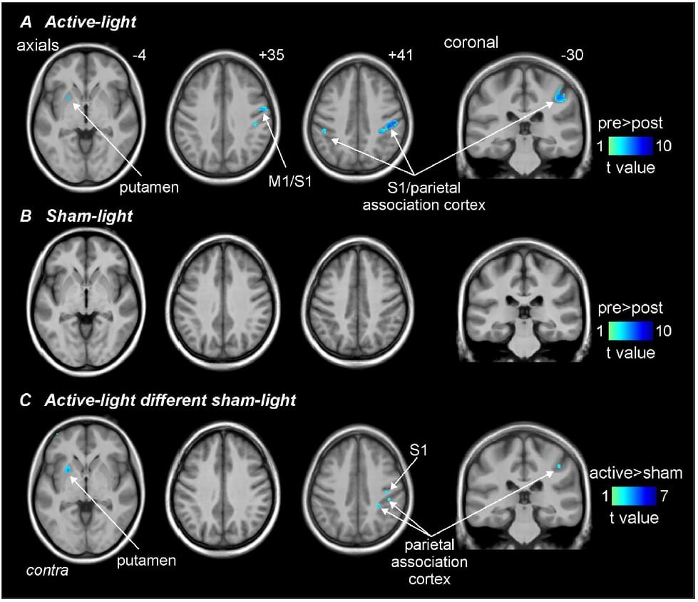
Effects of active and sham light on finger-tapping evoked activity. Source: El Khoury et al. (2019).
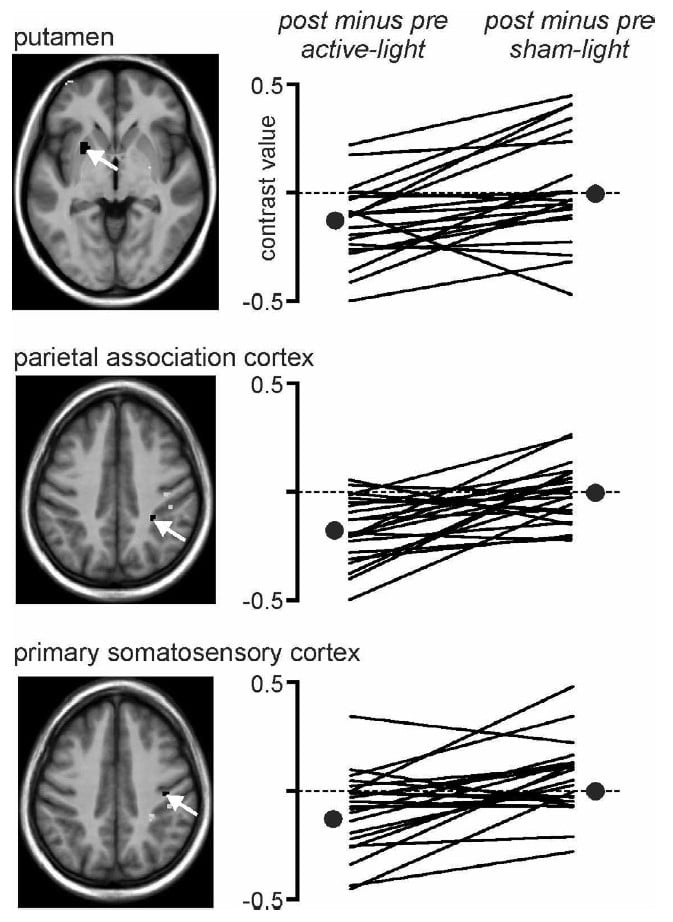
Individual participant activation under active and sham light. Source: El Khoury et al. (2019).
The dimmer switch vs. the precision dial
What people expect
Light acts like a dimmer switch and turns brain activity up across the board.
What the imaging suggested
At rest, nothing changed. When it was time to focus, background network activity got quieter. Less like a volume knob, more like a precision dial.
What this study does and doesn’t tell us
This was a single-session study in a small group of healthy young adults, and it measured brain activity patterns rather than performance. It doesn’t show long-term effects. It does offer an early look at how near-infrared light may interact with the networks behind everyday focus, and it gives future research a clear question to test.
Other neuroimaging research
The publications below used brain imaging in studies involving Vielight technology. They are listed for scientific reference only. Some studied populations or conditions for which Vielight devices are not intended, and listing them is not a claim that Vielight devices diagnose, treat, cure, mitigate, or prevent any disease. Vielight provided devices for, or contributed authors to, some of these studies.
Chao, L. L. (2019)
University of California San Francisco and San Francisco VA Medical Center. Arterial spin labeling perfusion MRI and resting-state fMRI. Photobiomodulation, Photomedicine, and Laser Surgery, 37(3), 133-141. doi:10.1089/photob.2018.4555
Chao, L. L., Barlow, C., Karimpoor, M., & Lim, L. (2020)
University of California San Francisco. Single-participant case report using structural MRI, resting-state fMRI, and perfusion MRI. Frontiers in Neurology. doi:10.3389/fneur.2020.00952
Wilde, E. A., et al. (2025)
University of Utah. Resting-state fMRI of cerebellar network connectivity. Photobiomodulation, Photomedicine, and Laser Surgery. doi:10.1177/25785478251376477
Wilde, E. A., et al. (2026)
University of Utah. Diffusion MRI in collegiate athletes over a competitive season. Journal of Neurotrauma. doi:10.1177/08977151251403554
Rashidi-Ranjbar, N., et al. (2025)
University of Toronto and St. Michael’s Hospital. Structural and functional MRI. Conference abstract. doi:10.1002/alz.095049
Explore our published research library
Vielight devices are general wellness products. They are not intended to diagnose, treat, cure, mitigate, or prevent any disease or condition, including Alzheimer’s disease, dementia, TBI, concussion, or CTE. Research findings described here are not medical advice.


